 |
| NanoDrop Spectrophotometer nucleic acid report of the boiling protocol, QuickExtract commercial kit, microwave protocol and enzymatic digestion followed by heating protocol. |
Thursday, March 30, 2017
NanoDrop Spectrophotometer Nucleic Acid Report
During this week, I focused primarily on the enzymatic digestion followed by heating protocol. Specifically, since this protocol has three different ways to perform it (using Ready-Lyse Lysozyme, proteinase-K, 10% Tween 20 and 20% SDS), I did not use Ready-Lyse Lysozyme and worked with the remaining substances. I was able to perform three sets of Eppendorf test tubes that used proteinase-K and another three that used proteinase-K plus 10% Tween 20 and 20% SDS. After I was done with the protocols, I ran them under the NanoDrop Spectrophotometer and the results are shown below. Not only does the graph show the values for these six test tubes but it shows the results of all of the protocols I have performed so far (boiling, QuickExtract commercial kit and microwave).

Thursday, March 23, 2017
Enterococcus faecalis
Enterococcus faecalis is a gram-positive opportunistic pathogen that generally causes urinary tract infections, wound infections and a variety of nosocomial (originate in a hospital) infections. To better see this bacteria, the first image shown below shows a Scanning Electron Micrograph (SEM) of it. Through fecal contamination, this bacteria is commonly found in food, sewage, water and soil (Paulsen, 2003). In other words, it can be found in a variety of materials we come into contact everyday. In addition, E. faecalis is able to withstand “oxidative stress, desiccation, and extremes of temperature and pH, and it has high endogenous resistance to salinity, bile acids, detergents, and antimicrobials” (Paulsen, 2003). Because of this, it has acquired “intrinsic resistance to many antibiotics and a remarkable capacity for developing resistance to others” and “now ranks among the most troublesome hospital pathogens” (Phillips, 1995). For example, E. faecalis has become resistant to vancomycin. The bacteria's genome carries its ability for resistance, which is shown below in the second image. As a result of its resistance, this bacteria “persist[s] in the hospital environment and survive[s] many host defenses” (McBride, 2007). Not only has it become resistant to various antibiotics and antimicrobial agents but it produces a two-subunit toxin, also known as cytolysin; this toxin is active against eukaryotic and prokaryotic cells. With the production of cytolysin, this bacteria is able “to actively probe the environment for target cells, and when target cells are detected, allows the organism to express high levels of cytolysin in response” (Coburn, 2004). However, this toxin is not the only material produced by E. faecalis, it also creates a “gelatinase, enterococcal surface protein Esp, aggregation substance, a hyaluronidase, and a bile salt hydrolase”, which are “known or suspected of enhancing the virulence” of this bacteria. (McBride, 2007).
Unfortunately, due to how complex this bacteria is, knowledge as to how it causes diseases and “transfer[s] antibiotic resistance to other pathogens” is limited (Nallapareddy, 2005). Therefore, because of its complexity, expanding knowledge on Enterococcus faecalis is evidently necessary and critical. In other words, being able to “characterize the genetic background of a [E. faecalis] strain makes possible the study of the flow of mobile elements within the species” (McBride, 2007). Being able to successfully accomplish this will “define the diversity of the E. faecalis species and to determine the core genome content” (McBride, 2007). With this, pharmaceutical scientists will be able to develop or improve drugs currently used on this bacteria and, thus, allow affected individuals to recover quickly and more efficiently.
Through DNA extractions of Enterococcus faecalis, identifying it, diagnosing infections and progressing in drugs will easily be achieved. Hence, in an attempt to quickly, successfully and effectively identify E. faecalis, four different DNA extraction protocols will be performed. These methods will determine which one is successful in yielding the most amount of DNA. The DNA extraction protocols are as follows: QuickExtract Bacterial DNA Extraction Kit, boiling method, microwave method and enzymatic digestion followed by heating method. These four procedures will be compared to a commercially produced kit. It is hypothesized that the boiling method will yield the most amount of DNA in Enterococcus faecalis. Following the boiling method, it is expected that the procedure to produce the second highest amount of DNA in E. faecalis will be the enzymatic digestion followed by heating. Lastly, the microwave method is expected to yield the least amount of DNA of this bacteria. In order to determine the quantity of DNA yielded from each extraction protocol, nanodrop spectrophotometry and electrophoresis will be performed.
Sources
Ahmed, O., Asghar, A., & Elhassan, M. (2014). African Journal of Microbiology Research. Comparison of three DNA extraction methods for polymerase chain reaction (PCR) analysis of bacterial genomic DNA. Retrieved February 23, 2017, from http://www.academicjournals.org/article/article1391446214_Ahmed%20et%20al.pdf
Coburn, P. S. (2004). Biography in Context. Enterococcus faecalis senses target cells and in response expresses cytolysin, 306. Retrieved March 23, 2017.
Dashti, A., Jadaon, M., Abdulsamad, A., & Dashti, H. (2009). Heat Treatment of Bacteria: A Simple Method of DNA Extraction for Molecular Techniques. Kuwait Medical Journal. Retrieved from https://www.researchgate.net/publication/266888615_Heat_Treatment_of_Bacteria_A_Simple_Method_of_DNA_Extraction_for_Molecular_Techniques
Englen, M., & Kelley, L. (2000). Letters in Applied Microbiology . A rapid DNA isolation procedure for the identification of Campylobacter jejuni by the polymerase chain reaction. Retrieved February 23, 2017, from http://onlinelibrary.wiley.com/store/10.1046/j.1365-2672.2000.00841.x/asset/j.1365-2672.2000.00841.x.pdf?v=1&t=izj0lngs&s=ffc6e6e746158600322301279b959afb27079119
Epicentre. (2012). QuickExtract™ Bacterial DNA Extraction Kit. Retrieved February 28, 2017, from http://www.epibio.com/docs/default-source/protocols/quickextract-bacterial-dna-extraction-kit.pdf?sfvrsn=8
McBride, S., Fischetti, V., LeBlanc, D., Moellering, R., & Gilmore, M. (2007). PLOS One. Genetic Diversity among Enterococcus faecalis. doi:10.1371/journal.pone.0000582
Nallapareddy, S., Wenxiang, H., Weinstock, G. M., & Murray, B. E. (2005). Journal of Bacteriology. Molecular Characterization of a Widespread, Pathogenic, and Antibiotic Resistance-Receptive Enterococcus faecalis Lineage and Dissemination of Its Putative Pathogenicity Island. Retrieved March 23, 2017.
Oliveira, C. F., Paim, T. G., Reiter, K. C., Rieger, A., & D'azevedo, P. A. (2014, Jan. & feb.). Evaluation of four different DNA extraction methods in coagulase-negative staphylococci clinical isolates. Retrieved February 23, 2017, from https://www.ncbi.nlm.nih.gov/pmc/articles/PMC4085835/
Paulsen, T., Banerjei, L., & Myers, G. S. (2003). Science. Role of Mobile DNA in the Evolution of Vancomycin-Resistant Enterococcus faecalis. doi:10.1126/science.1080613
Phillips, D. M., Ph.D. (1995). Images in Clinical Medicine (K. Eagle M.D., Ed.). Enterococcus faecalis. doi:10.1056/NEJM199501053320105
 |
| Scanning Electron Micrograph (SEM) of Enterococcus faecalis. (Phillips, 1995). |
 |
| Enterococcus faecalis genome (Paulsen, 2003). |
Thursday, March 9, 2017
NanoDrop Spectrophotometer & EMCC Research Abstract
During my fourth week, I did not continue to test any of my DNA extraction methods. However, with the methods (QuickExtract, boiling and enzymatic digestion followed by heating using Ready-Lyse Lysozyme) that I performed last week, I was able to figure out how much DNA was extracted from Enterococcus faecalis using a NanoDrop spectrophotometer (image shown below). Even though I do not know how to read the graphs and they all look different, I hope I correctly performed them. As mentioned earlier, I was not in the lab throughout the week. Instead, I was busy writing and editing my Estrella Mountain Community College (EMCC) research abstract for the student conference. I am looking forward towards presenting my project, even though I am a little nervous.
 |
| NanoDrop Spectrophotometer |
Thursday, March 2, 2017
DNA Extraction Methods on Enterococcus Faecalis (Week 3)
During my 3rd week, I began working on my research project, which involved 4 DNA extraction methods following a set of protocols. I am trying to determine which DNA extraction method will yield the most of amount of DNA from Enterococcus faecalis. The methods that I am testing include a commercial kit and 3 (one includes 3 different chemicals to use) methods that I chose. The name of the commercial kit is QuickExtract. For this method, I followed a protocol and used QuickExtract Bacterial DNA and Ready-Lyse Lysozyme (1st image shown below). Since it is a commercial kit, it is supposed to yield the most amount of DNA. As for the other 3 methods, I will use protocols for a boiling, microwave and enzymatic digestion followed by heating (done using 3 different chemicals) method. The boiling method did not require any chemicals so I only needed my bacteria, a centrifuge and a water bath. For the microwave method, I will need a centrifuge, TE buffer (2nd image shown below), 15% SDS, a microwave, lysis buffer and ethanol. As for the enzymatic digestion followed by heating method, a centrifuge is needed along with 3 different chemicals (only that each time I perform the protocol, only one chemical is used). The 3 different chemicals that I will use are Ready-Lyse Lysozyme, proteinase K, 10% Tween 20, and 15% SDS (proteinase K, 10% Tween 20, and 15% SDS will be used altogether in during one protocol). However, during this week, I was only able to perform 3 methods: QuickExtract, boiling and one of the ways to perform enzymatic digestion followed by heating (I used Ready-Lyse Lysozyme). Next week, I will be working on the microwave method and using the remaining chemicals for the enzymatic digestion followed by heating method.
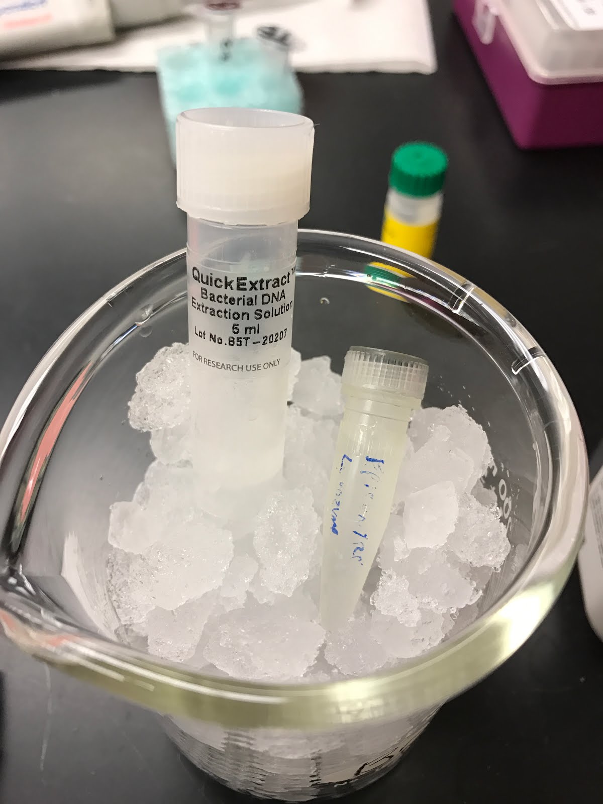 |
| QuickExtract Bacterial DNA & Ready-Lyse Lysozyme |
 |
| TE buffer |
Thursday, February 23, 2017
Gram+ Cocci Tests
During my 2nd week of the S-STEM program, I worked more with my unknown bacteria, enterococcus faecalis. I performed 6 different tests on it to verify that I indeed identified the bacteria correctly. These tests involved glucose fermentation, lactose, tryptone, TSI, gelatin, TSB and a MSA plate (respectively, as shown below). However, the 1st MSA plate that I streaked on Tuesday, February 21, 2017 did not grow any colonies so I streaked another plate. Still, 2 days later, both of these plates did not show any changes. Nonetheless, the other tests worked as expected. For the glucose fermentation, before the 24 hours (as shown below), the color of glucose was orange and the durham tube (glass tube inside the glucose tube) did not have any air bubbles, which meant a gas was produced. However, over a day, the color changed to yellow and the durham tube still did not have an air bubble (as shown below). This meant that my bacteria produced an acid and did not produce gas. In addition, the lactose tube also changed color from pink to yellow and had no air bubble. Again, the change in color and no air bubble present in the durham tube meant that my bacteria produced an acid and did not produce a gas. To continue, for the tryptone test, I added indole reagent to test if indole was present in my bacteria. If the top layer turned red, indole was present but if the top layer turned yellow, indole was not present. In this case, indole was not present because the top layer of tryptone is yellow (shown below in the 2nd image). As for the TSI tube, I made an “s” shape on the inside using a loop that was dipped into my bacteria. As a result, since it did not turn black, it was negative for H2S (hydrogen sulfide) gas. Similar to the previous tests, the TSI tube turned yellow, which also meant an acid was produced. Next, the gelatin tested whether my bacteria had gelatin hydrolysis. After the 24 hours, the gelatin melted and was now a liquid. To test if it had gelatin hydrolysis, I put the gelatin in the fridge for 10 minutes. If it went back into its solid form, it was negative but if it stayed as a liquid, it was positive. For my bacteria, after the 10 minutes, it went back into its solid form and, thus, was negative for gelatin hydrolysis. Finally, the TSB tube ended up looking cloudy, which meant that bacteria successfully grew. All in all, the 6 tests that I performed showed a change over a course of a day, except for the MSA plates.
 |
| 6 gram+ cocci tests before 24 hours |
 |
| 6 gram+ cocci tests after 24 hours |
Thursday, February 16, 2017
STEM Beginnings
I started my internship on Monday, February 13, 2017. In my first week, I was introduced to the basics. For example, I watched safety videos, learned how to clock in/out, received my lab notebook, began working in the lab and identified an unknown bacteria. In order to identify my unknown, I used a plastic loop to streak it onto a TSA plate (top image). Once I was done with that, I left it in a Biohazard fridge for 24 hours. The next day, colonies (the small circles) of bacteria were successfully formed (2nd image). In order to identify my bacteria, I used these colonies to perform gram staining and a catalase test. When working on gram staining, the chemicals used were crystal violet, iodine, alcohol decolorizer and safranin. The purpose of the gram staining was to determine if it were gram+ or gram- (if it were purple, it would be gram+; if it were pink, it would be gram-). Some portion of my bacteria had both pink and purple present, but, since the majority of it was purple, I concluded that it was gram+ (3rd image). Not only did I have to find if it was gram+ or gram-, but I had to figure out if it were coccus (circle) or bacillus (rod) shaped. After closely examining the bacteria, I determined that it was coccus (bottom image). After that, I did a catalase test, which involved using a drop of hydrogen peroxide. Basically, after adding the drop, if it bubbled, I had to follow other tests to ID my bacteria, but, luckily, it did not bubble so I identified it as Enterococcus faecalis. All in all, my first week was a new experience and I cannot wait to see what this semester holds!
 |
| TSA plate before 24 hours. |
 |
| TSA plate after 24 hours. |
 |
| Unknown bacteria after gram staining. |
 |
| Enterococcus faecalis (gram+ & coccus shaped). |


Subscribe to:
Posts (Atom)